It‘s Ursula’s third time in the functional MRI machine. Heather Kosakowski, a PhD student in cognitive neuroscience, is hoping to get just two precious minutes of data from her session. Even though Ursula has been booked to have her brain scanned for two hours, it’s far from a sure thing. Her first two sessions, also booked for two hours each, yielded only eight minutes of usable material combined.
The task verges on the impossible. Kosakowski needs Ursula to stay both awake, watching projected images of faces and scenes, and very still, ideally for seconds or even minutes at a time. Every twitch and wiggle blurs the MRI’s scan, obscuring the image and rendering it useless. But Ursula tends to squirm and then, inevitably, fall asleep—exactly what you would expect from a six-month-old baby.
Scanning infants’ brains while they’re awake is incredibly difficult and time consuming, and there’s always a risk that a session will produce no data at all. A motivated adult is capable of staying perfectly still for two hours, yielding brain images that read like an open book. Ursula’s sessions produce something more akin to a book that’s been torn up and thrown in a river. Kosakowski, who is jointly advised by cognitive neuroscience professors Nancy Kanwisher ’80, PhD ’86, and Rebecca Saxe, PhD ’03, needs to carefully fish out the usable sections and stitch them together before she can read the story they reveal.
“Heather is probably the most skilled person alive today at getting high-quality functional MRI data from human infants,” says Kanwisher, the Walter A. Rosenblith Professor of Cognitive Neuroscience.
If Kosakowski doesn’t get the two minutes of still brain images she needs, it’s possible Ursula’s two previous visits will have been for nothing. But if she can get a clear fMRI reading, she will be one step closer to answering one of the most profound questions in modern neuroscience: What are the physical underpinnings of the human mind?
A far-off dream
Kosakowski is no stranger to overcoming obstacles. Her childhood was characterized by the sort of instability that made any kind of higher education feel unattainable. Her father was in the military, so her family moved around a lot. Then her parents divorced, and the difficulties multiplied. When she was about seven, she and her mother moved into a homeless shelter, and at 11, Kosakowski was put into foster care in Western Massachusetts. “Having a bachelor’s degree was always my dream,” she says, but she calls her first attempt at college a “dismal failure.”

“I dropped out, I was homeless and had no job, and then I totaled my car and I was like, what am I going to do with my life?” she recalls. She decided to enter the Marine Corps but hoped she might someday get a second shot at college.
After several years in the military, Kosakowski left the Marines and returned to Massachusetts, enrolling part time at Massachusetts Bay Community College. Maybe, she thought, a bachelor’s degree wasn’t so far out of reach after all. She set her sights on Smith College, where she was admitted. But around the time she received that piece of news, she also got another: she was pregnant.
Kosakowski didn’t go to college anywhere that fall, but her eagerness to learn persisted. Her natural curiosity found an outlet in her baby, Hannah, who was born that October. She watched Hannah experience grass for the first time in the spring, delighting at how she reacted to this strange new material covering the ground. As Hannah explored her environment, Kosakowski kept wondering what things looked like from her perspective. What was she experiencing? How was she making sense of the world around her?
When Hannah was two, Kosakowski got a job at a nonprofit that was working to speed up research on multiple sclerosis, giving her the opportunity to attend a neuroscience conference. “After that I was kind of hooked,” she says. “I was like, okay, the thing I really want to be doing is research, and in order to do research I need a degree. I have to go back to school.”
Kosakowski was accepted to Wellesley College—a school she had written off years before because it was so competitive. She became fascinated with neuroscience, peppering her professors with questions until one said, “Heather, some of the questions you ask, nobody knows the answer. You should get a PhD.”
“I just kept scanning”
Just as Kosakowski graduated from Wellesley, Saxe was looking for a manager for her lab, which happened to be one of the only labs in the world studying babies in MRIs while they are awake—and consequently one of the only ones that could answer Kosakowski’s questions about the nature of infant cognition. She applied for the position and got it.
When her foster sister had a baby, Kosakowski went to Saxe and asked if she could learn how to use the fMRI to scan the infant’s brain. “I think she thought I’d just scan my niece and be done with it,” Kosakowski says. “But I just kept scanning, and she never told me to stop.” Saxe, impressed with Kosakowski’s work and determination, agreed to take her on as a graduate student. In 2017, she left her job as lab manager and began her first semester as a PhD candidate at MIT.
Kanwisher still remembers the first time she saw Kosakowski scan a baby in the MRI. She was considering working with Kosakowski and Saxe on their infant study, but at first she was highly skeptical. “This kid is squealing like crazy. The mom is nervous. The whole thing is stressful. And this goes on and on and Heather’s not giving up,” she says. “Then—boom. The clouds part, the kid smiles, Heather pops the kid into the MRI scanner, and like a minute later she scans beautiful data. Just the persistence, the skill, is spectacular.”
Kosakowski’s current study focuses on how the brains of babies just two to nine months old respond to short videos of faces or bodies—and how that differs from their response to scenes without people. She is the first to find evidence of this kind of specialization in children under five years old. The information she’s after can only be measured with fMRI. Magnetic resonance imaging allows researchers to take high-resolution pictures of cross-sections of the brain. Functional MRI adds another layer to this, recording images of brain activity in real time. When neurons in one section of the brain are particularly active, blood flow increases to fuel that region. This shows up as a bright spot on fMRI scans.
Researchers have spent decades using fMRI to prove that sections of the adult brain are highly specialized for certain tasks, and to pinpoint exactly which areas are specialized for which functions. “The mind and brain have all this structure. We’re not just generically smart. We’re smart in very particular ways about very particular things that humans do,” Kanwisher says. “If you look at the structure, you see this set of dozens of regions of the brain, each that does a very distinctive, different thing … It’s impossible to look at that and not wonder, ‘How did that structure get wired up?’”
When adults look at faces, for instance, a section of the brain called the fusiform face area, or FFA, lights up. In other words, if researchers put adults in MRI machines and show them images of both faces and objects, the FFA will only respond to faces. The parahippocampal place area (PPA), meanwhile, responds most strongly to depictions of scenes. Kanwisher herself named the FFA after leading the team that discovered it in 1997, and she led the effort to pinpoint and describe the PPA in 1998. She and her colleagues also discovered the brain section known as the extrastriate body area (EBA), which responds strongly to pictures of body parts, in 2001.
“[Heather is] scanning the youngest awake humans that anybody can scan and asking what structure is in the brain within a few months of birth,” Kanwisher says. “And that’s just one of the most thrilling questions in all of psychology, neuroscience, and deep philosophy: What is the structure of our minds and where does it come from?”
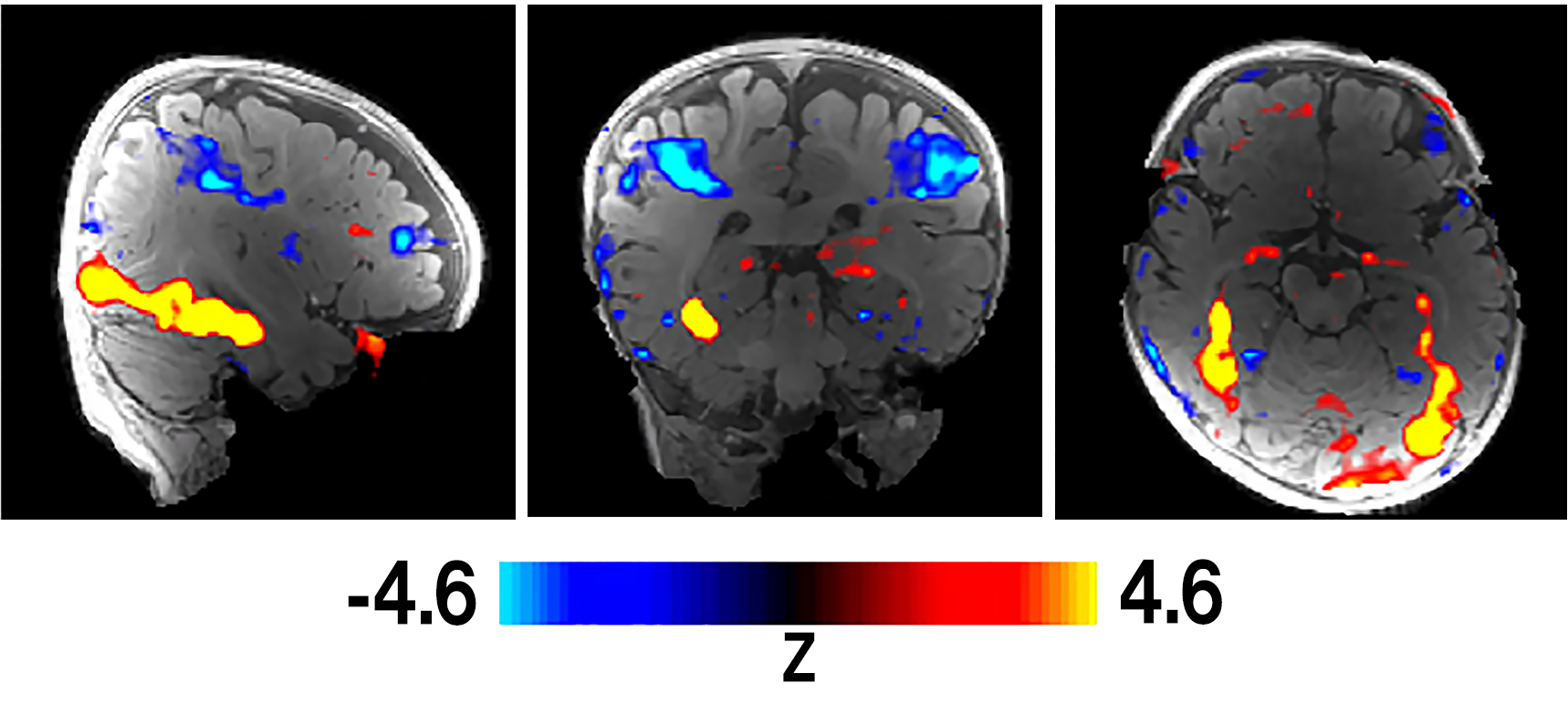
Preliminary results from Kosakowski’s study provide some of the strongest evidence yet that some functions of our brain may be innate rather than learned. “Babies also have selective responses for faces, bodies, and scenes in the FFA, EBA, and PPA,” Kosakowski says. “Nobody has ever found that before. And it was completely not expected that we would find that.”

watching videos of faces and scenes, red and yellow show activity related to viewing faces and blue shows activity linked to scenes.
The covid-19 pandemic, though, has brought its own set of challenges. The fMRI sessions had to be put on hold, and she has spent much of the last year analyzing her data at home. “Covid-19 has greatly impacted me in that I am working from home full time and also a single parent—the same way it’s impacted lots of families with children,” Kosakowski says. She hopes to have the opportunity to scan more infants to begin studying their auditory processing before graduating in May 2022. But Ursula’s session in late 2019 turns out to have been one of her last chances to get precious data for her dissertation.
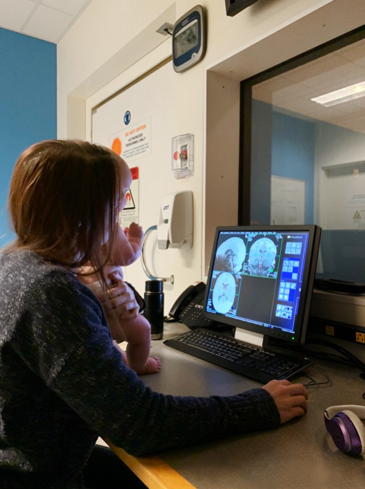
In that session, Ursula had fallen asleep in the scanner, but Kosakowski was in high spirits as it wrapped up. She was pretty sure she’d managed to get the data—and she’d later confirm she was right. As Ursula’s mother left to change out of MRI-friendly scrubs, Kosakowski scooped up the groggy baby and held her as she sat in front of a computer. The unflappable grad student has an uncanny ability to keep babies happy and relaxed—a skill no PhD program will teach you, but one that’s invaluable when every extra second of clean data helps.
Finally, she pulled up the image she’d been looking for, pointing toward the screen as Ursula’s gaze followed her finger. Someday, thanks to their combined efforts, we might have a better understanding of how Ursula perceived the image before her. “Look!” Kosakowski told the baby in her arms. “That’s your brain!”
